
Our sense of vision is one of the incredible ways we can explore and connect with our surroundings. And, taking care of our eyes should be a top priority, like other areas of our health and wellness.
Luckily, technological advancements give us a simple and noninvasive approach to better understanding areas of our health we cannot otherwise see, no matter how far back we roll our eyes.
Optomap is a retinal imaging tool that provides a wide-view snapshot of your eye’s internal structures. This makes it easier to detect potential issues early and track any changes over time.
Routine eye exams offer a comprehensive insight into your vision and eye health, and Optomap helps illustrate this full picture.
All About Optomap
The retina is a thin layer of light-sensitive tissue lining the back of the eye and plays a central role in your visual and ocular health. It’s responsible for sensing incoming light entering the eye and converting it into electrical signals, allowing us to see clear, sharp images.
A comprehensive view of your eye health requires assessing internal structures like the retina, and advancements like Optomap make this easier than ever.
Optomap retinal imaging captures up to 82% of your retina in a single panoramic shot, surpassing standard methods that cover only about 15%, allowing for easier detection of issues in the retina’s peripheral areas.
This wide-field imaging allows your optometrist to gain a deeper understanding of your retinal health in just seconds, making it a valuable tool for preserving your visual and ocular health. Read the stories of patients who used an Optomap machine during an eye exam.
How Does Optomap Work?
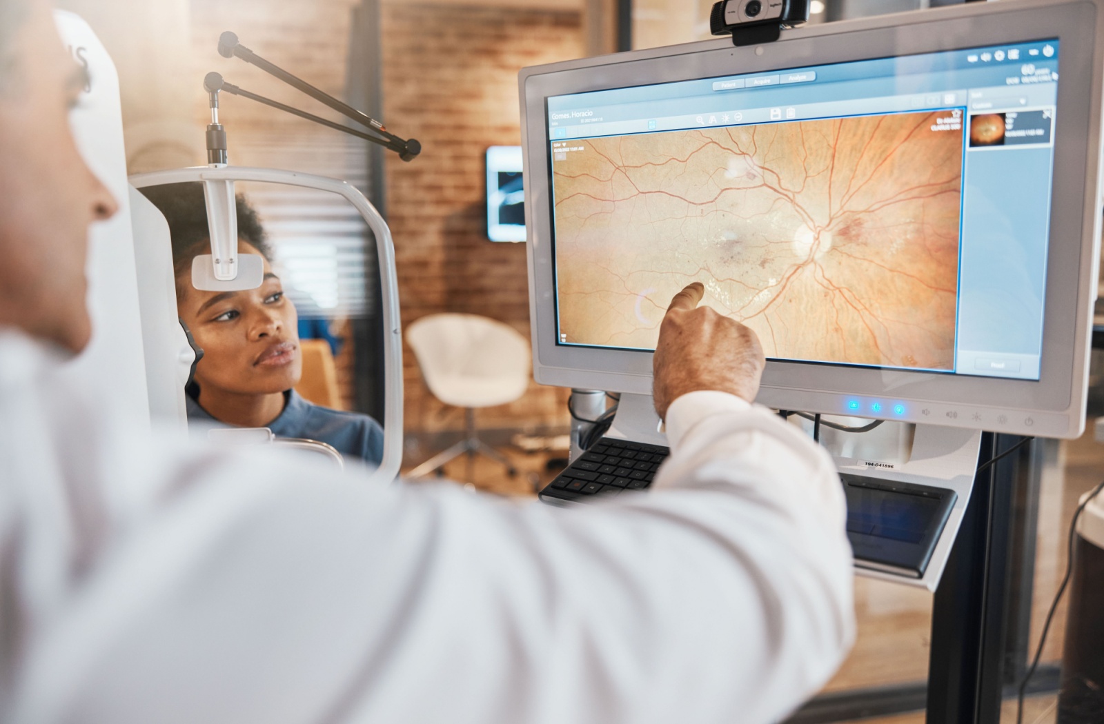
Optomap uses scanning laser technology to capture images. Two low-power lasers (one red and one green) scan the back of your eye to produce a detailed, high-resolution image of the retina.
These images illustrate a snapshot of your current retinal health, and over time, these images serve as a benchmark for tracking any changes.
The process is quick, straightforward, and does not require a dilation. With the touch of a button, your eye doctor obtains valuable data that can identify the earliest signs of diseases such as glaucoma, macular degeneration, and diabetic retinopathy.
Optomap’s low-power lasers are safe, gentle, and suitable for almost everyone.

Image of an eye that previously had a retinal detachment: taken by an Optomap machine.
How Retinal Imaging Supports Eye Health
Retinal imaging involves capturing detailed snapshots of the retina to assess its health and detect abnormalities. This tool allows eye doctors to identify eye and general health conditions.
These detailed images can be taken during different types of eye exams, including:
- Routine Eye Exams: To establish a baseline measurement of your retina and monitor its health over time.
- Dilated Eye Exams: If your optometrist suspects more significant issues, a dilation may be used to widen the pupils, to gain a closer look at the eye’s internal structures.
- Emergency Visits: When you experience sudden symptoms such as flashes of light, floaters, or vision loss, retinal images help detect any abnormal changes to the eye’s internal structures.
Retinal images serve as a diagnostic tool for detecting the earliest stages of eye conditions, preventing progression, and protecting your vision.
- Glaucoma: Increased eye pressure can cause peripheral vision loss. Damage or cupping of the optic nerve in retinal images may indicate the presence of glaucoma.
- Diabetic Retinopathy: A diabetes-related complication that affects the blood vessels in the retina. Small blood vessel leaks, retinal swelling, or growth of abnormal blood vessels in retinal images may indicate the presence of diabetic retinopathy.
- Age-related Macular Degeneration (AMD): A progressive condition that can result in central vision loss. Signs that suggest the presence of this condition include drusen deposits beneath the retina, pigmentary changes in the macula, and progressive thinning or scarring of the macula.

Image of an eye with a floater: taken by an Optomap machine.
Flashes and Floaters Found With Retinal Imaging
Flashes appear as brief streaks of light in your field of vision. They often happen unexpectedly, even in a completely dark room or with your eyes closed. Meanwhile, floaters appear as small shadowy shapes drifting across your visual field, often resembling squiggly lines or cobwebs.
Floaters appear more commonly with age, as the vitreous gel becomes more liquid, clumping tiny fibers together, which casts shadows on your retina, creating the sensation of something “floating” in your vision.
Flashes occur when the vitreous pulls or tugs on the retina, stimulating its light-sensitive cells. This tug creates the illusion of light, even though no external light source is present.
Though flashes can vary in frequency and intensity, they’re not typically “normal”. Similarly, while floaters may be harmless, experiencing new floaters suddenly or in large quantities may suggest an underlying concern worth looking into.
Any damage or disruptions to your retina, which is responsible for processing visual input, are detrimental to your sight. The onset of flashes or changes in floaters may indicate serious eye conditions, like retinal detachments or tears. When left untreated, these conditions can lead to permanent vision loss.
Therefore, it’s imperative to visit your eye doctor as soon as you notice new symptoms related to flashes or floaters, such as:
- A sudden increase in floaters or newly visible floaters
- Bright, recurring flashes of light
- A shadow or “curtain” creeping across your vision
- Blurred vision or loss of central or peripheral vision
Fortunately, Optomap’s precision and wide-field imaging can help identify the underlying cause of these visual disruptions by capturing detailed images of any abnormal or structural retinal changes. With timely intervention and the click of a button, we can preserve your overall health and vision.
Say Cheese: Taking Your Retinal Photos
Optomap is non-invasive, gentle, and quick, but for those wondering what to expect during this scan, here is a quick step-by-step overview to ease any hesitation before your next eye exam:
- Align your chin and forehead: You will be asked to rest your chin on a chin rest and place your forehead gently against a support bar. This helps stabilize your head for the imaging process.
- Focus on a target: Similar to having your photo taken, you will be asked to focus on a target. Your eye doctor or technician will ask you to look at a specific point of light inside the imaging device. This verifies your eyes are positioned correctly for an accurate scan.
- Your scan is taken: A low-power laser scans your retina. The process is entirely painless and takes just seconds.
- Review your results: Once the image is captured, your doctor will immediately review the scan with you. This is a great opportunity to better understand your retinal health and discuss any findings.
- Done and dusted: That is it! No downtime, no discomfort, and no dilation necessary (unless your optometrist would like to investigate further). You are all set for clearer insights into your eye health.
Invest in Your Vision with Optomap
Your eyes are invaluable, and taking steps to preserve their health is one of the greatest long-term investments you can make for yourself. Optomap sets a strong foundation for proactive vision care by capturing detailed images of your retinal health. Connect with our Hawkesbury Optometric Clinic team to schedule your next routine eye exam to see your eyes up close and personal!